


University of Michigan School of Dentistry faculty member helps develop new recommendations for significant change related to dental X-rays8 min read
Ann Arbor, Mich., March 5, 2024 – A University of Michigan School of Dentistry faculty member helped develop a new recommendation by the American Dental Association (ADA) that is likely to affect millions of Americans during their future dental appointments.
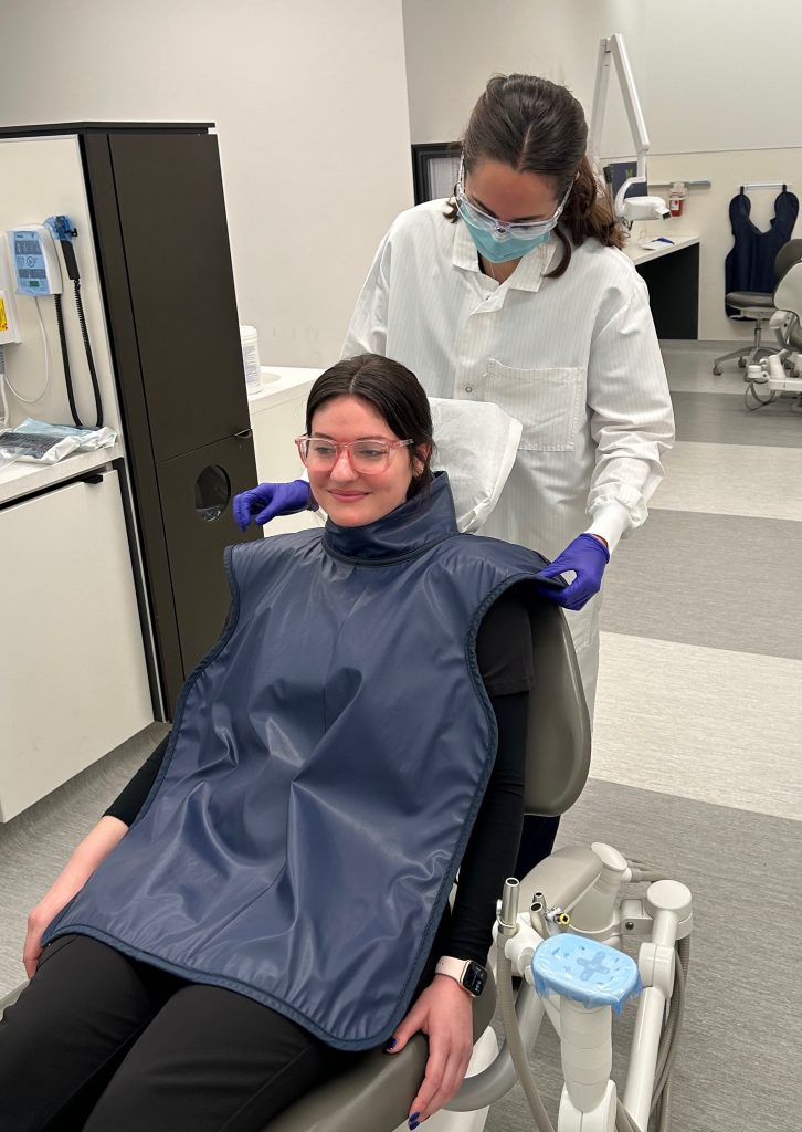
Dr. Erika Benavides was chair of an expert panel established by the ADA’s Council on Scientific Affairs to review the long-standing recommendation that lead aprons and thyroid collars should be used to shield dental patients from radiation exposure during X-ray procedures.
Working with ADA staff over the last three years, Benavides and other radiology experts from around the country reviewed the latest published research, documents and regulations regarding radiation exposure. The review determined that lead aprons and thyroid collars are not necessary to protect patients from harmful levels of radiation, leading the ADA to recommend the end of a practice that has been standard procedure for decades. The recommendation applies to all patients, including pregnant women and children, and all types of intraoral and extraoral dentomaxillofacial imaging, including Cone-Beam Computed Tomography (CBCT).
The practice of lead shielding in dental offices was implemented in the 1950s based on indirect evidence and concerns that exposure to large or repeated doses of radiation could cause cancer, damage reproductive organs or harm a fetus. In addition to large lead aprons covering the patient’s chest and abdomen area, thyroid collars are often placed around the patient’s neck because the thyroid gland, located on the neck close to the oral cavity, is particularly sensitive to radiation as a cause of thyroid cancer.
However, the recent ADA study on dental imaging safety and radiation protection determined that the use of lead aprons and thyroid collars is unnecessary because of advances in radiology technology and the lack of evidence that the low levels of radiation produced during dentomaxillofacial imaging can cause heritable effects or thyroid cancer. The latest digital imaging technology, the use of a more-focused X-ray beam, and the appropriate patient-based selection of needed radiographs and exposure factors have been found to be significantly more effective in decreasing the radiation dose.
Dr. Benavides, a Clinical Professor in the Department of Periodontics and Oral Medicine at the U-M School of Dentistry, co-directs and teaches numerous introductory and advanced didactic and clinical radiology courses for dental, dental hygiene and graduate students. She is a Diplomate and Past-President of the American Board of Oral and Maxillofacial Radiology (ABOMR) and has served as Councilor for Communications and Chair of the Research and Technology Committee of the American Academy of Oral and Maxillofacial Radiology (AAOMR).
Benavides said the ADA review involved the collaboration of ADA researchers, the expert panel and consultants from various dental specialties, such as endodontics, periodontics, orthodontics, oral surgery, oral pathology, and pediatric dentistry. Participants met in a series of online meetings as they reviewed data and research, then formulated recommendations and collaborated on the study that was published in the Journal of the American Dental Association (JADA).
Medical physicists with the U.S. Food and Drug Administration (FDA) supported the panel’s development of these recommendations. They are also aligned with recent recommendations released by the AAOMR based on a review committee that included Benavides.
The ADA process was thorough and rewarding, Benavides said. The participants reached a consensus that not only changed the longstanding patient shielding recommendations, but also included additional ways that dentists and their staff members can make radiology as safe as possible. That’s important, the study noted, because radiographic imaging procedures used in dental practices are collectively among the most frequently performed in healthy people in the United States, as well as one of the most common radiographic examinations performed worldwide. The goal of the study, Benavides said, is to provide up-to-date information to help clinicians implement the latest safety protocols that provide the best diagnostic benefits while minimizing radiation risks for patients and dental office staff.
She said an important reminder for dentists and their staff members in the report is that radiographs should be ordered based on the patient’s needs, for specific diagnostic purposes. “Dentists need to order radiographs based on the patient’s clinical exam findings and not just at set intervals,” Benavides said. “Taking ‘routine bitewings’ at every dental cleaning appointment is not good practice, for example. The interval at which recall bitewing radiographs are prescribed should be customized for each individual patient based on their caries and periodontal disease risk and clinical findings.”
It is unlikely that dental patients around the country will notice immediate changes during their next dental appointment, Benavides said, because dentists will need to study the new recommendations, develop new protocols, train their staff, and educate their patients. Also, dentists are bound by various federal regulations governing the use of radiation; some states have legislation as well. Benavides said the ADA and professional radiology organizations will engage federal and state regulators in an educational effort about the new ADA recommendation to encourage uniform application of radiology procedures.
Benavides said that the state of Michigan, for example, does not have regulations mandating the use of the patient shielding, but that doesn’t necessarily mean the dental school or private dental practices in the state will immediately drop the use of lead aprons and thyroid collars. Each practitioner will need to consider the best way to adapt to the new recommendation based on their individual radiation safety protocols. At the dental school, with an existing curriculum already including radiology standards, implementation of the new recommendation will likely take a couple of months as all faculty, students and staff need to be trained and educational materials for patients need to be developed.
The new study also reports that the use of shields can actually increase radiation exposure instead of protecting against it. That’s because positioning a shield, particularly the thyroid collar, may inadvertently obstruct the area to be X-rayed if it is not precisely placed. That can require repositioning it and taking a second image, thus increasing the exposure. Similarly, lead shields can interfere with the automatic settings of digital computed tomography scanners, which take a preliminary scout view to determine the optimal exposure factors; if the preliminary scan reads the dense lead shield, it may set the dosage higher than it needs to be, which would result in an increased radiation exposure to the patient. Additionally, the research determined that the lead shields have negligible impact on preventing radiation from scattering away from the target site into the rest of the body.
Benavides noted that radiology education and updates are advancing in multiple areas of healthcare, providing additional affirmation for the new ADA recommendation. The university’s health system, Michigan Medicine, began a “Shed the Shield” campaign at the end of last year for diagnostic radiographs and scans of different parts of the body including the abdomen, which typically require higher doses of radiation than dental x-rays.
Another U-M School of Dentistry faculty member, Dr. Purnima Kumar, is chair of the ADA Council on Scientific Affairs, which commissioned the report. Dr. Kumar said one of the key takeaways of the report is the need to avoid unnecessary X-rays. “The central point of these recommendations is that clinicians should order radiographs in moderation to minimize both patients’ and dental professionals’ exposure to ionizing radiation,” said Kumar, professor of dentistry and chair of the Department of Periodontology and Oral Medicine. “We encourage dentists and their teams to review these best-practice recommendations, comply with radiation protection regulations and talk with their patients about any questions or concerns before ordering dental imaging.”
The ADA is the nation’s largest dental association, representing 159,000 dentist members. The organization has advocated for the public’s health and promoted the art and science of dentistry since 1859.
###
The University of Michigan School of Dentistry is one of the nation’s leading dental schools engaged in oral healthcare education, research, patient care and community service. General dental care clinics and specialty clinics providing advanced treatment enable the school to offer dental services and programs to patients throughout Michigan. Classroom and clinic instruction prepare future dentists, dental specialists and dental hygienists for practice in private offices, hospitals, academia and public agencies. Research seeks to discover and apply new knowledge that can help patients worldwide. For more information about the School of Dentistry, visit us on the Web at: www.dent.umich.edu. Contact: Lynn Monson, associate director of communications, at [email protected], or (734) 615-1971.