
Digital Imaging Advancing Care at School of Dentistry5 min read
Ann Arbor, MI — October 2, 2013 — Digital imaging is making significant inroads at the University of Michigan School of Dentistry.
New digital imaging equipment was recently installed in 22 clinics throughout the School to enhance the quality of care patients receive as well as prepare students in dental, dental hygiene, and graduate programs for the digital environment they will experience after graduation.
The latest efforts build upon the successful use of digital technology in the Dr. Roy Roberts Preclinical Laboratory as well as the digital impression system available in dental student clinics.
For 10 years, clinical instructors have been using digital technology to demonstrate dental procedures that first- and second-year dental students watch on a monitor at each of 110 workstations. Students apply that knowledge and develop their clinical skills on models of the oral cavity (typodonts), plastic teeth, and mannequin heads prior to treating patients in the School’s clinics. More recently, students have been taking digital impressions and replacing traditional stone models with highly accurate and durable stereolithography models.
Benefits Cited
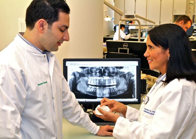
The new digital imaging equipment now being used includes intraoral X-ray units, sensors and phosphorous plates, as well as panoramic, panoramic-cephalogram, and cone beam computed tomography machines that provide excellent visualization of the teeth and bony structures of the head.
The radiographic images “will appear on a monitor in seven seconds or less,” said Roger Gillie, director of Application Services in Dental Informatics.
Students and clinical faculty can zoom in on a digital image, highlight details and share radiographs. For example, residents in the oral surgery clinic, he said, “will be able to see radiographs taken in the emergency or predoctoral clinics before the patient is taken to that clinic for evaluation and treatment.” Electronic enhancements allow for greater review of image details and notes can be added as reminders of areas to watch in the future.
Light boxes that have been used for many years to view film X-rays have been removed in the radiology clinic. However, in four clinics where predoctoral students treat patients under the supervision of clinical faculty, light boxes will remain since it will take about a year to scan tens of thousands of film radiographs to digital format.
Dr. Stephen Stefanac, associate dean for Patient Services, said, “the quality of the digital X-ray images is exceptional and should help us to detect problems in patients earlier. Also important,” he added, “is that students can now show detailed images to their patients to help them understand the treatment options that are available.”
Extensive Rollout Preparation
The digital imaging initiative involved extensive collaboration among clinicians, radiologists, and staff in Patient Services, Radiology, Dental Informatics and others throughout the School, according to Dr. Erika Benavides, clinical assistant professor and oral and maxillofacial radiologist. She led the clinical digital imaging team.
“It was a giant research project,” Benavides said with a smile. “We reviewed a considerable amount of information from many different hardware vendors and, because the equipment had different capabilities, evaluated each system using objective criteria.”
Also critical was choosing the digital imaging software to use. “The software is the foundation that allows us to capture and store the images using the different radiology equipment, and allows clinicians and students to share the images throughout the School or even across campus,” she said. “Our clinician users were actively involved in evaluating the software, so their input was essential in making a final decision.”
As Benavides talked to faculty and students, Gillie led a team of 12 people who “looked under the hood” to assess the digital technology being considered and evaluate its effectiveness. “The images had to be shared among 22 clinics at the School and three others at U-M Hospital where the School of Dentistry also has a clinic. The technology also had to be robust and user friendly for students, dental clinical faculty, students in graduate dental programs, and in research. Every area was important,” he said.
The Paper to Digital Transition
As the new school year began, Terri Stilwell, radiology clinic instructor, trained small groups of dental, dental hygiene, and graduate students how to use the new equipment. “The students are adapting to the changes very well and are excited to use the new technology,” she said. “They’re tech savvy and amazed at how easy it is to use.”
The transition to digital imaging has also led to a change in vocabulary.
As she trained one group of predoctoral students, Stilwell caught herself saying “film X-rays” and quickly corrected that. “It’s hard to break an old habit, especially when one has used a phrase like ‘film X-rays’ for so many years,” she said with a smile. Let’s use ‘digital images’ instead.”
After January 1, 2014, the School will no longer create a paper record for new patients. “We will then digitize portions of 30,000 existing records,” Stefanac said. “When the paper records are in digital form and combined with new digital images, our students will have easier access to all their patient information.”
Eventually, digital images will be accessible on Apple and Android mobile devices. All information will be encrypted to comply with all protected health information standards.
Collaboration Across U-M
“It was a great team effort. People from many disciplines came together to make this happen,” Benavides said. “Our faculty members were excited to learn about the capabilities of the new digital equipment and how to use it. Dental students were also enthused because they use technology extensively.”
Gillie lauded the help received from the School’s Technology Services group, U-M’s Information Technology Communications (ITCOM) unit and the Medical School’s radiology department. “Working together we installed 80 imaging stations that allow us to take, store and manage digital images that are available on more than 1,000 computers,” he said. “Digital imaging is an excellent example of how the School of Dentistry is using technology to enhance the educational experience of students and the care provided to their patients.”